1. What is EEG?
Similar to the electrocardiogram (ECG) and electromyography (EMG), electroencephalography (EEG) is a non-invasive method for recording the brain's electrical activity. As shown in the figure below, during an EEG measurement, electrode sensors are attached to the scalp. These sensors transmit the synchronized activity of neurons to a computer, which reflects the extracellular currents. It is important to note that EEG primarily reflects the activity of cortical neurons near the brain electrodes, while deep structures such as the hippocampus, thalamus, and brainstem do not directly influence the EEG.
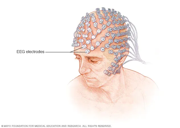 ▷ EEG apparatus. Source: mayoclinic
EEG enables the detection of various brain wave frequencies and can be used to assess different forms of sleep and epilepsy. In a normal human EEG, brain activity primarily ranges from 1 to 30 Hz. The main types of brain waves include α waves (8-13 Hz), β waves (13-30 Hz), δ waves (0.5-4 Hz), and θ waves (4-7 Hz).
α waves: These are typical of a relaxed, wakeful state and are most prominent in the parietal and occipital regions. For example, when the brain is in a state of relaxation or meditation, it enters the α wave state. β waves: These low-amplitude waves become more prominent during intense neural activity, primarily appearing in the frontal lobe and other regions, and are considered a hallmark of working memory and logical thinking. θ and δ waves: These waves occur during drowsiness and early sleep stages. Their presence in an awake state may indicate brain dysfunction.

▷ Various brain wave patterns. Source: infoinstruments
2. The Birth in 1924
As early as 1755, Italian scientists Luigi Galvani, Alessandro Volta, Georg Ohm, and Michael Faraday demonstrated the remarkable electrical properties of biological tissues.
In 1870, Gustav Theodor Fritsch conducted studies on a dog's brain and published an article titled On the Electrical Excitability of the Brain. He discovered the electrical excitability of the exposed cerebral hemispheres in the dog's brain, concluding that certain areas of the brain were responsible for motor functions. However, his research was met with great skepticism in the academic community, since it was widely believed that electrical currents were generated deep within the brain rather than in the cortex.
In 1913, Russian physiologist Prawdwicz-Neminski recorded EEGs from a dog. Due to the extremely small electrical changes detected by the electrodes and the lack of amplification techniques, the experiment had to be conducted on an exposed brain. Prawdwicz-Neminski was the first to describe a 12 to 14 Hz rhythm under normal conditions and coined the term "electroencephalogram."
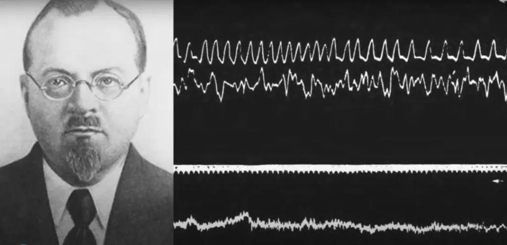 ▷ Prawdwicz-Neminski. Source: The Brainclinics Foundation / YouTube
It was the German psychiatrist Hans Berger who truly introduced EEG to the scientific community. Berger’s interest in EEG research stemmed from his fascination with telepathy involving his sister. In 1893, at the age of 19, Berger fell from his horse during a German military exercise and was nearly trampled. On that same day, his sister, who was far away, had an ominous premonition about him. She convinced their father to send a telegram asking if Hans was alright. For the young Berger, this eerie coincidence was not merely chance but a case of “spontaneous telepathy.” Convinced that he had transmitted his fearful thoughts to his sister, Berger decided to study psychiatry in an effort to uncover the mystery of how thoughts could be transmitted between people. This serendipitous event led Berger to make a significant contribution to modern medicine and science: he invented the EEG and was the first to systematically record EEGs from the human brain.
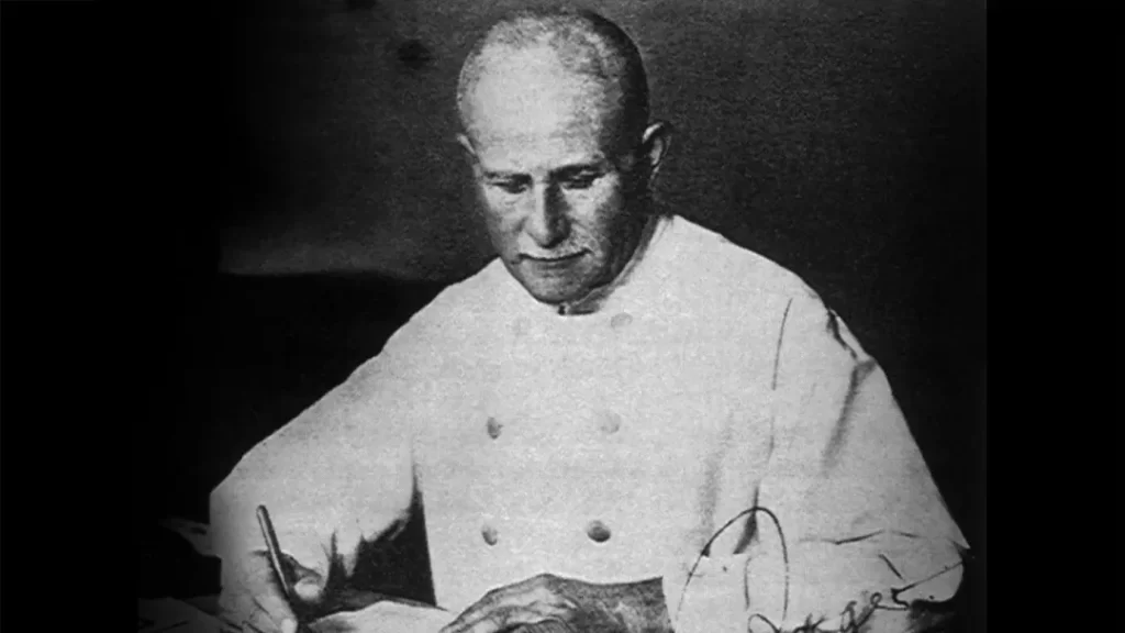 ▷ Hans Berger. Source: sciencenews
In July 1924, a 17-year-old boy underwent neurosurgery, during which part of his brain was exposed due to a head injury. Taking advantage of this unexpected opportunity, Dr. Hans Berger recorded the boy’s EEG through the exposed cranial window. Following this, Berger began attempting to record EEGs through the scalp, with his first volunteer being his son. Using a simple single-channel device, he successfully depicted α rhythms and β activity, despite the recordings being brief, lasting only 1 to 3 minutes. He identified the normal occipital background rhythm and the faster frequency β waves, which marked the official birth of EEG. Berger eventually accumulated 1,133 recordings from 76 patients. Despite publishing 14 scientific reports, he did not receive the recognition he deserved at the time, partly because his papers were written in German.
Throughout his research on human EEGs, Berger kept pace with the latest technological advancements. He used various devices, starting with the initial Einstein model, then the Edelmann, and eventually the more powerful Siemens model. In 1929, he used the Siemens instrument to record the human EEG traces, which he reported in his first publication.
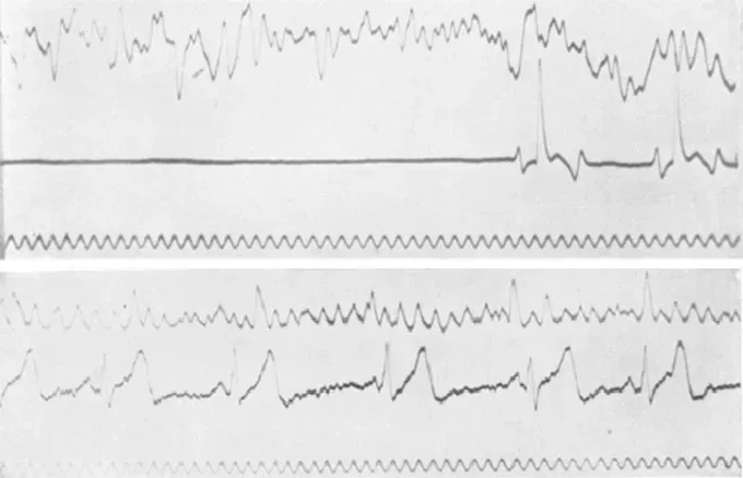 ▷ The first human EEG results published by Hans Berger. Source: sciencenews
By 1934, British scientists Lord Edgar Adrian and Brian Matthews had verified Berger’s findings, known as the "Berger Rhythm," in a public demonstration at the Cambridge Physiological Society, later publishing their results in the journal Brain. With the technical improvements made by Adrian and Matthews, Berger continued his research, discovering that the widespread and regular brain rhythms changed when subjects opened their eyes or engaged in problem-solving.
Throughout the 1930s, Berger continued his studies on human EEGs, publishing several reports that included some valuable findings, such as research on consciousness fluctuations, sleep spindles, the effects of hypoxia on the human brain, various diffuse and localized brain diseases, and even some indications of epileptic discharges.
Tragically, the later years of this great scientist’s life took a dark turn. Before the outbreak of World War II, he was expelled from his research position at the University of Jena in Germany and forced to take non-research work in a nursing home. Believing he was suffering from a fatal heart disease and afflicted with infections and depression, he ultimately took his own life in 1941. The scientific community now agrees that Berger deserved a Nobel Prize in Physiology or Medicine, but by the time he was nominated in 1942 and 1947, the founder of EEG had long passed away.
3. From Europe to North America
Around 1935, the emerging field of electroencephalography (EEG) began to shift its focus from Europe to North America, led by a group of researchers who crossed the Atlantic. On this new stage, American EEG science rapidly gained international recognition, particularly through the research of Hallowell Davis at Harvard University, Frederic A. Gibbs and Erna L. Gibbs, and Herbert H. Jasper at Brown University.
A. J. Derbyshire, a graduate student under Davis, was deeply inspired by Berger's 1929 paper and had been attempting to demonstrate the α rhythm, but his efforts repeatedly failed. It wasn't until 1934 that Davis recorded his own α rhythm, sparking great excitement in the laboratory.
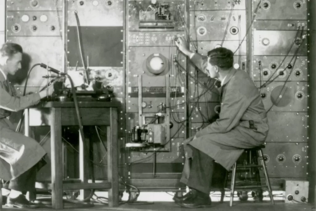 ▷ A. J. Derbyshire (left), Hallowell Davis, and early EEG equipment. Source: oto.wustl.edu
That same year, a breakthrough in clinical EEG was achieved in the study of epilepsy patients. Frederic A. Gibbs established a connection with William G. Lennox, a prominent epileptologist of the time. Lennox had already begun studying cerebral circulation by measuring the oxygen and carbon dioxide content in jugular vein blood. Erna L. Gibbs, an immigrant from Germany, was Lennox's technical collaborator and later became his wife. A self-taught EEG technician, Erna Gibbs became one of the world's earliest EEG technicians and contributed to many important research papers, including a landmark study on cerebral blood flow.
In the subsequent years, EEG gained more attention than cerebral blood flow in Lennox's research. Notably, Lennox, the Gibbses, and Davis collaborated on a study that identified the association of 3 Hz spike-wave complexes with petit mal absences in 12 epileptic children. This finding has since become a standard diagnostic tool in EEG.
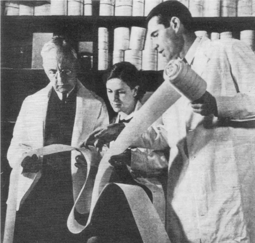 ▷ In 1937, William G. Lennox, Erna L. Gibbs, and Frederic A. Gibbs examining EEG traces on paper rolls. Source: Stone, James L., and John R. Hughes. "Early history of electroencephalography and establishment of the American Clinical Neurophysiology Society." Journal of Clinical Neurophysiology 30.1 (2013): 28-44.
Unlike Berger, who was fascinated by oscillatory rhythms, Frederic Gibbs was more interested in paroxysmal EEG patterns, such as spike waves. The Gibbses and Lennox later reported on the EEG patterns associated with grand mal seizures and psychomotor epilepsy. However, the rapid spike-wave intervals in grand mal seizures and the 4 or 6 Hz rhythmic activity in psychomotor epilepsy did not garner as much attention as the 3 Hz spike-wave pattern in petit mal epilepsy.
Recognizing the limitations of EEG recording techniques, the Gibbses visited Hans Berger in Germany in the summer of 1935 and studied the "polyscope" instrument of J. F. Toennies at the Berlin Buch Research Institute. During their time in Europe, they also came across the EEG instrument developed by Matthews in England. Frederic Gibbs later contacted Albert Grass at MIT to manufacture a three-channel preamplifier, resulting in the Grass Model I EEG. This device included three channels and an ink-writing recorder, which captured the traces on paper rolls. The Grass Model I came into use in 1935, marking the beginning of the commercial EEG era.
Looking back, the 1930s era of Gibbs-Gibbs-Lennox proved to be one of the most exciting and productive periods in EEG history. During those years, EEG demonstrated its greatest utility in the study of epileptic seizure disorders.
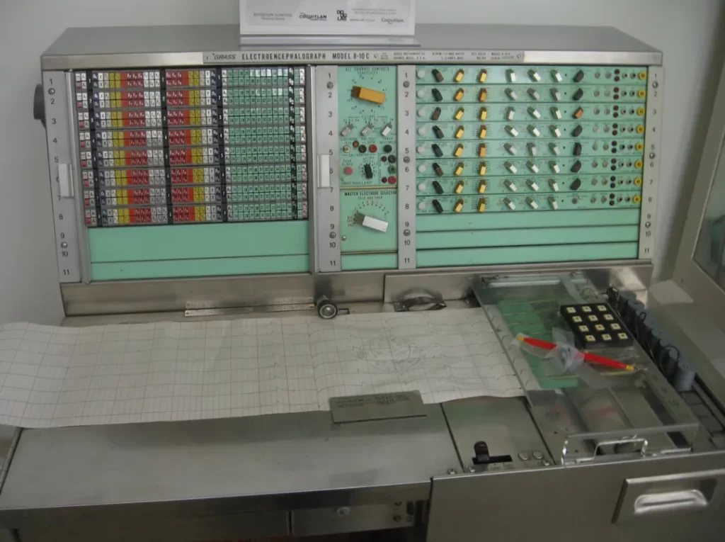 ▷ Grass Model 8-10C EEG machine. Source: Niall Williams
Another noteworthy pioneer in clinical EEG research during the 1930s was William Grey Walter. Walter's work in 1936 established him as a trailblazer in clinical EEG. He was the first to apply EEG in the diagnosis of brain tumors and to monitor the effects of anesthesia during surgery. Walter identified δ and θ waves in slow brain activity and introduced the first method for automatic analysis of brain bioelectrical signals.
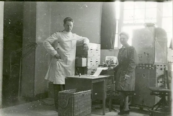 ▷ William Grey Walter (left). Source: Wikipedia
From 1960 onwards, EEG became a clinical tool not only for studying and diagnosing epilepsy and space-occupying lesions but also as a valuable diagnostic and prognostic test in many other medical fields. At the same time, EEG contributed to defining and standardizing various sleep stages and provided valuable information for the study of sleep disorders.
However, during World War II (1939-1945), research and clinical EEG activities were restricted, especially in Europe. After the war, the gap between North American and European EEG research widened significantly, with European efforts falling into decline. Meanwhile, across the Atlantic, Frederic Gibbs and his colleagues conducted a significant study on anterior temporal lobe spike discharges during the interictal period in patients with psychomotor epilepsy, an important work in elucidating the mechanisms of temporal lobe epilepsy that had far-reaching implications for the development of EEG research.
Despite his international reputation and recognition as a leader in clinical EEG, Gibbs's status at Harvard did not match his stature. He held only a lecturer's post, with status and salary far below that of a professor, even though many of his European peers longed to visit his laboratory.
4. The Clinical Adoption of EEG
In the 1950s, EEG became widely recognized in the medical field. In the early 1950s, almost every academic tertiary care center had at least one EEG machine. By the late 1950s, EEG equipment had spread to many smaller hospitals and even private clinics. Central and departmental EEG laboratories were established in university hospitals, and the field of pediatric EEG also began to develop.
Some psychiatric departments took particular pride in their clinical and research-oriented EEG work, as doctors had already recognized that many diseases affecting the central nervous system could be detected by corresponding signs on an EEG.
Neurosurgeons like Wilder G. Penfield and A. Earl Walker, who were focused on epilepsy research, began to show interest in EEG and its application on the cortical surface or within deeper cortical structures. By the late 1950s, researchers such as Meyers, Hayne, and Knott pioneered the use of implanted intracranial electrodes for deep EEG monitoring in humans. In the 1960s, deep EEG became essential in the surgical treatment of intractable epilepsy.
Following 30 years of rapid and sustained development in clinical and experimental EEG research, the field reached its peak around 1960, after which research activity began to decline. EEG experts in academic institutions shifted their focus from tracing brain wave patterns to automated data analysis. Notably, Cooley and Tukey introduced the Fast Fourier Transform (FFT) in 1965 to extract the spectral or frequency content of signals, laying the foundation for power spectral analysis. Their work set new standards for EEG computerization, paving the way for the quantitative EEG (qEEG) we use today.
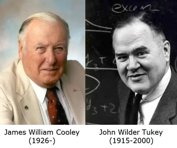 In the 1970s and 1980s, the advent of structural neuroimaging technologies like computed tomography (CT) and magnetic resonance imaging (MRI) appeared to challenge EEG’s role in diagnosing structural brain abnormalities. However, this shift was more superficial than substantive. EEG was never intended as a structural test; it remains the only diagnostic tool capable of providing large-scale, real-time measurements of neocortical dynamic function.
Later, topical EEG diagnosis re-entered the spotlight in the form of computerized brain function localization. This development is closely associated with the name Frank Duffy, whose work Clinical Electroencephalography and Topographic Brain Mapping reinvigorated EEG technology, achieving an innovative blend of traditional techniques with modern advancements.
5. Outlook and Conclusion
Today, EEG is utilized both clinically and in research to further elucidate brain function. Its developmental trajectory can be summarized as becoming "spatially finer and temporally more precise." In modern neuroscience research, EEG is used to study human attention, consciousness, language, and decision-making processes. Researchers aim to gain deeper insights into the workings of the brain by analyzing the dynamic characteristics of brain waves.
Clinically, beyond its traditional applications in epilepsy and sleep studies, EEG traces hold the potential to reveal further clues to neurological disorders. For instance, combining EEG biomarkers with AI could facilitate early detection, diagnosis, and monitoring of treatment in various diseases. Additionally, the development of brain-computer interface technology has introduced new applications for EEG. These include aiding severely disabled individuals in communication and helping paralyzed patients regain motor function through prosthetics.
What will EEG be like in the next century? We cannot know for certain. However, if EEG recording becomes as convenient as tracking heart rate with a fitness watch, its applications could expand significantly. At that point, EEG could transcend research and clinical boundaries, becoming an integral part of everyday life for all.